About Macular Holes
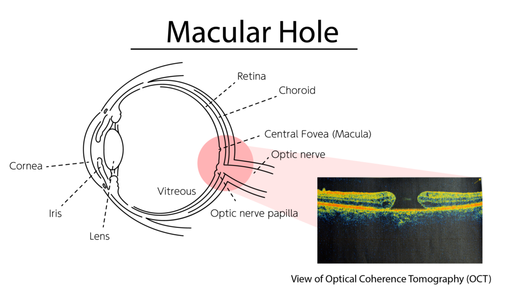
The eye essentially functions like a camera with a lens that focuses light through the clear vitreous gel that fills the eye and onto the retina. The retina is inside the eye (near the back of the eye) and functions like the film in a camera. The retina processes what we see and sends a signal to the brain for further interpretation. As we get older, the vitreous gel in the eye becomes liquefied and separates from the retina. In a small percentage of people, as the vitreous gel pulls away from the retina, it tugs (or causes traction) on the macula. The macula is the part of our retina which is responsible for clear, sharp vision and allows us to read small print and appreciate fine details. Occasionally a membrane may also form on the retina which can also cause traction (tugging) on the macula. Excessive traction can ultimately lead to a hole forming in the macula. This is termed a macular hole.
Macular Hole Symptoms
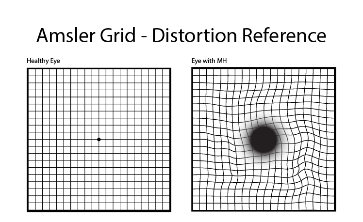
A macular hole can cause blurred and distorted central vision. Sometimes straight lines can appear bent or wavy. Macular holes are related to aging and usually occur in people over age 60. Macular holes and age-related macular degeneration are two separate and distinct conditions, although the symptoms for each can be similar. Without treatment, the blurry vision caused by a macular hole usually worsens to an ultimate visual acuity of 20/400 (the size of the big “E” on the eye chart) or worse.
Diagnosis and Tests for Macular Holes
In order to diagnose a Macular Hole, your retinal specialist will have to perform imaging or an optical coherence tomography in order to get a better view of the retina. y.
Macular Hole Surgery
RCA & The AAO Present Retina Macular Hole Surgery
Prior to the early 1990s, there was no available treatment for macular holes. Since the discovery of macular hole surgery, anatomic success with closure of the macular hole can be achieved in over 90% of most cases. With hole closure, most patients notice improvement in their vision, although the vision does not become “perfect.” The surgery involves removal of the vitreous gel (vitrectomy) and replaced with a temporary gas bubble. After the surgery, patients are required to maintain a face-down position to allow the gas bubble to press against the hole, allowing it to close itself. The gas bubble will slowly be absorbed by the body over a period of 2-8 weeks, depending on the type and concentration of gas this used by your surgeon. The eye does not make more vitreous gel after it is removed. Instead, the eye replaces the gas bubble with a watery fluid. While the gas bubble is in the eye, you cannot travel to higher elevations, fly in an airplane, or have specific types of anesthesia, such as nitrous oxide (“laughing gas;” commonly used by dentists). Any of these activities can lead to severe pain and possible blindness. Your doctor will tell you when it is safe to resume these activities.
To assist with the face-down positioning, we can provide you with information regarding positioning aids that may be rented from specialty companies. Ask your doctor for this information.
Minimal Or “Non” Face-Down Positioning
There is extensive literature, including papers published by RCA physicians, which show an excellent success rate with minimal face-down positioning. Many macular holes can be closed without any face down positioning at all, and these can be identified during a dilated exam with non-invasive imaging. The success rate is not compromised. Patients must keep in mind, however, that altitude restrictions remain until the gas is fully dissolved. To view physicians who do not require face down posturing, click here.
As with any surgery, there are risks involved. These risks include cataract formation, retinal tear or detachment, failure of hole closure, and infection or bleeding in the eye. If you have not already had cataract surgery, you will likely develop a cataract and may need cataract surgery in the next 6-12 months. A retinal detachment occurs in 1-2 percent of patients undergoing macular hole surgery, and it is usually repairable. With these relatively low risks, many patients elect to proceed with surgery with a realistic expectation of improved vision when the surgery is performed in a timely fashion.
Here is a great tool for patients who plan to travel or adjust elevations after having surgery – Elevation Calculator – please consult your physician before traveling.
FAQs
Can a retinal hole fix itself?
It is rare for a macular hole to go away on it’s own. Most often, the best treatment option is to perform surgery.
Can a macular hole cause blindness?
A macular hole, when left untreated can drastically affect your central vision. This the vision that the macula is responsible for. It does not, however, affect your peripheral vision.
How do you recover from macular hole surgery?
After the surgery, patients are required to maintain a face-down position to allow the gas bubble to press against the hole, allowing it to close itself.
Hear what patients are saying
Other Conditions Treated at RCA
Our ophthalmologists are experienced in diagnosing and treating many retinal conditions.


